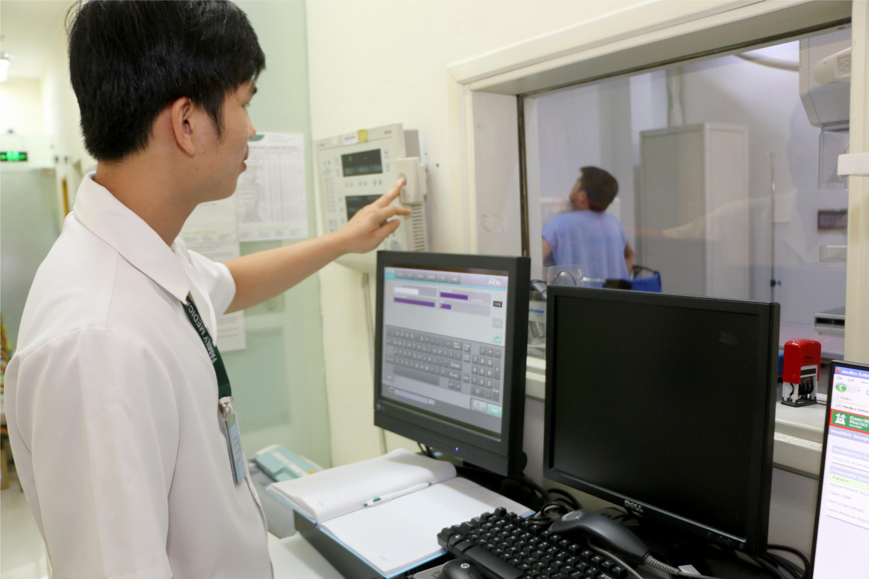
Chẩn đoán hình ảnh

Khoa chẩn đoán hình ảnh tại Care24H cung cấp một loạt các dịch vụ chẩn đoán và chẩn đoán hình ảnh hỗ trợ cho cả khoa cấp cứu và ngoại trú của chúng tôi.
Thời gian chờ đợi cho tất cả các dịch vụ chẩn đoán rất ngắn do quyết định của Care24H cung cấp cho tất cả bệnh nhân dịch vụ theo dõi nhanh.
Phần lớn các dịch vụ của chúng tôi là tại nhà và có sẵn 24 giờ một ngày.
Bác sĩ X quang, kỹ thuật viên và bác sĩ siêu âm Care24H có nhiều năm kinh nghiệm và chuyên môn tuyệt vời trong lĩnh vực họ chọn.
Các bộ phận hình ảnh cung cấp các dịch vụ sau:
X-quang hoặc X quang nói chung
Các hình thức y tế thường được sử dụng nhất. Tia X có thể tạo ra hình ảnh chẩn đoán cơ thể người bằng kỹ thuật số trên màn hình máy tính.
Chụp X-quang cực kỳ nhanh và cung cấp một phương pháp nhanh chóng để đánh giá toàn bộ cơ thể, đặc biệt là khớp, xương và ngực. Xương X-quang xương xác định và điều trị gãy xương bao gồm cánh tay, chân, đầu gối, cổ tay, vai, cột sống và hộp sọ.
X-quang ngực thường được thực hiện để đánh giá phổi, tim và thành ngực trong điều trị viêm phổi, suy tim, khí phế thũng, ung thư phổi và các điều kiện y tế khác.
Sê-ri Đường tiêu hóa trên / dưới (GI) Hữu ích để kiểm tra các vết loét, khối u lành tính (polyp) hoặc các dấu hiệu của một số bệnh đường ruột khác, ví dụ: Bệnh Crohn.
Chụp cắt lớp CT
Kết hợp một loạt các hình ảnh X quang được chụp từ các góc khác nhau và sử dụng xử lý máy tính để tạo ra các hình ảnh cắt ngang hoặc lát cắt của xương, mạch máu và các mô mềm bên trong cơ thể bạn. Hình ảnh quét CT cung cấp thông tin chi tiết hơn so với tia X thông thường. Quét CT có thể được sử dụng để hình dung gần như tất cả các bộ phận của cơ thể và được sử dụng để chẩn đoán bệnh hoặc chấn thương, cũng như lên kế hoạch điều trị y tế, phẫu thuật hoặc xạ trị.
MRI Scanning sử dụng sóng từ trường và sóng vô tuyến mạnh để tạo ra hình ảnh trên máy tính của các mô, cơ quan và các cấu trúc khác bên trong cơ thể bạn. MRI Scanning tạo ra hình ảnh rõ ràng của hầu hết các bộ phận của cơ thể. Điều này rất quan trọng khi tia X có thể cung cấp đủ thông tin cần thiết. Được sử dụng để có được hình ảnh chi tiết của não và tủy sống và để phát hiện các bất thường và khối u. Dây chằng bị rách xung quanh khớp có thể được phát hiện bằng quét MRI; nó đang được sử dụng ngày càng nhiều sau chấn thương thể thao.
Hình ảnh nhi
Máy quét CT đã giảm thiểu nhu cầu an thần ở trẻ em và cho phép giảm đáng kể liều phóng xạ. Những lý do phổ biến để chẩn đoán hình ảnh ở trẻ em: khối u Wilms, bệnh bạch cầu, quái thai, bất thường bẩm sinh, viêm xương, viêm màng não, hội chứng suy hô hấp ở trẻ sơ sinh, viêm khớp vô căn ở trẻ vị thành niên, gãy xương do thiếu máu xanh.
Chụp nhũ ảnh / hình ảnh vú
Quá trình sử dụng tia X năng lượng thấp để kiểm tra vú của con người. Mục tiêu của chụp nhũ ảnh là phát hiện sớm ung thư vú. Chụp ảnh vú được thực hiện kết hợp với chụp nhũ ảnh để bác sĩ có cái nhìn rõ ràng hơn về vú của con người. Mục tiêu của hình ảnh vú là phát hiện sớm ung thư vú.
Siêu âm tổng quát (3D / 4D)
Được sử dụng để hình dung cơ bắp, gân và nhiều cơ quan nội tạng; để ghi lại kích thước, cấu trúc của chúng và bất kỳ tổn thương bệnh lý nào bằng hình ảnh thời gian thực. Các xét nghiệm bao gồm bụng, thận, động mạch chủ, tuyến giáp, tuyến tiền liệt, tinh hoàn, sản khoa, phụ khoa và nhi khoa.
Siêu âm sản phụ khoa
Siêu âm sản khoa là việc sử dụng siêu âm trong thai kỳ. Có thể đánh giá các chuyển động như nhịp tim của thai nhi và dị tật ở thai nhi, và các phép đo có thể được thực hiện chính xác trên các hình ảnh hiển thị trên màn hình.
Siêu âm tim
Kiểm tra siêu âm sử dụng sóng âm thanh cường độ cao được gửi qua một thiết bị gọi là đầu dò. Thiết bị phát ra tiếng vang của sóng âm thanh khi chúng bật ra khỏi các phần khác nhau của trái tim bạn. Cung cấp thông tin, tức là kích thước và hình dạng của tim (định lượng kích thước buồng bên trong), khả năng bơm, vị trí và mức độ của bất kỳ tổn thương mô nào. Có thể cung cấp cho các bác sĩ ước tính chức năng tim như tính toán cung lượng tim, phân suất tống máu và chức năng tâm trương (tim thư giãn tốt như thế nào).
Nội soi dạ dày và nội soi
Một thử nghiệm kép được thực hiện với thuốc an thần nhẹ. Thử nghiệm đầu tiên để nhìn trực tiếp vào cổ họng, dạ dày và phần đầu tiên của bạn. Thử nghiệm thứ hai kiểm tra ruột lớn.

 We use cookies on this website to enhance your user experience